Electrospun cellulose acetate phthalate nanofibrous scaffolds fabricated using novel solvent combinations biocompatible for primary chondrocytes and neurons
Rupendra Shrestha 1 2*, Asha Palat 1 2*, Alan M Punnoose 2, Shailesh Joshi 3, D Ponraju 4, Solomon F D Paul 1
- Department of Human Genetics, Sri Ramachandra University, Chennai, India;
- Cell and Tissue Engineering Laboratory, Centre for Regenerative Medicine and Stem Cell Research, Sri Ramachandra University, Chennai, India.
- Radiological Safety Division, Indira Gandhi Centre for Atomic Research (IGCAR), Kalpakkam, India.
- Physical and Chemical Analysis Section, Safety Engineering Division, Indira Gandhi Centre for Atomic Research (IGCAR), Kalpakkam, India.
Publication: Tissue and Cell. 2016 Dec;48(6):634-643. doi: 10.1016/j.tice.2016.07.007
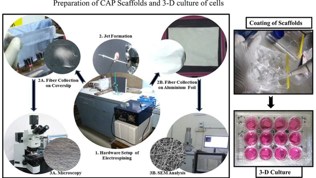
Abstract: Electrospun nanofibres have been shown to exhibit extracellular matrix (ECM)-like characteristics required for tissue engineering in terms of porosity, flexibility, fibre organization and strength. This study focuses on developing novel cellulose acetate phthalate (CAP) scaffolds by electrospinning for establishing 3-D chondrocyte and neuronal cultures. Five solvent combinations were employed in fabricating the fibres, namely, acetone/ethanol (9:1), dimethylformamide/tetrahydrofuran/acetone (3:3:4), tetrahydrofuran/acetone (1:1), tetrahydrofuran/ethanol (1:1) and chloroform/methanol (1:1). The electrospun fibres were characterized by scanning electron microscopy (SEM) analysis and confirmed to be within the nanometre range. Based on the morphology of the fibers from SEM results, two solvent combinations such as acetone/ethanol and dimethylformamide/tetrahydrofuran/acetone were selected for stabilization as CAP exhibits a pH dependent solubility. Fourier-Transform Infrared (FTIR) analysis revealed the hydrolysis of CAP which was overcome by EDC [1-ethyl-3-(3-dimethylaminopropyl) carbodiimide] and EDC/NHS (N-hydroxysuccinimide) cross-linking resulting in its stability (pH of 7.2) for three months. MTT [3-(4, 5-dimethylthiazol-2-yl)-1, 5-diphenyltetrazolium bromide] assay performed using L6 myoblast confirmed the biocompatibility of the scaffolds. 3-D primary chondrocyte and neuronal cultures were established on the scaffolds and maintained for a period of 10 days. H&E staining and SEM analysis showed the attachment of the chondrocytes and neurons on CAP scaffolds prepared using dimethylformamide/tetrahydrofuran/acetone and acetone/ethanol respectively.
Cite: Shrestha R, Palat A, Punnoose AM, Joshi S, Ponraju D, Paul SF. Electrospun cellulose acetate phthalate nanofibrous scaffolds fabricated using novel solvent combinations biocompatible for primary chondrocytes and neurons. Tissue Cell. 2016 Dec;48(6):634-643. PMID: 27546071